MS SPIDOC
Mass Spectrometry for Single-Particle Imaging of Dipole Oriented Protein Complexes.
The European XFEL has just entered user operation. With its unparalleled peak brilliance and repetition rate, European XFEL has the potential to further applications in single particle imaging (SPI), thus far limited to large viral particles at X-ray fee-electron lasers (XFEL). SPI will allow imaging protein complexes without the need for crystallization. This eventually renders transient conformational states accessible for high resolution structural studies, yielding molecular movies of biomolecular machines. A major bottleneck is the wealth of data required to reconstruct a single structure leading to long processing times. This is currently also a problem in electron microscopy (EM). MS SPIDOC will overcome this data challenge by developing a native mass spectrometry (MS) system for sample delivery called X-MS-I. It will provide mass- and conformation-selected biomolecules that are oriented along their dipole axis upon imaging. This will enable structural reconstruction from much smaller datasets, speeding up the analysis time tremendously. Moreover, the system features low sample consumption and a controlled low background, easing pattern identification.

The Main Objectives Of The Project Are:
- Deliver mass and conformation separated biomolecules for SPI
- Orient proteins for SPI
- Image protein complex unfolding
- Exploit potential of protein orientation for other applications.
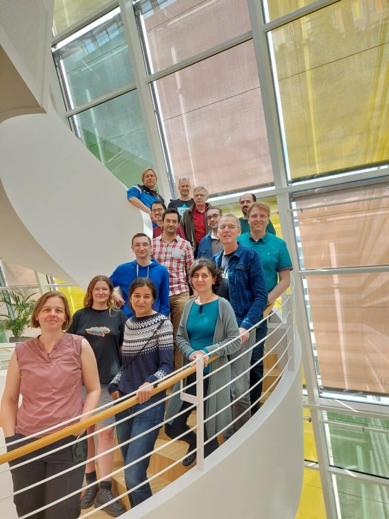
The MS SPIDOC consortium combines internationally leading expertise in different fields relevant to the project: Instrument design and development, computer simulations as well as working with biomolecules in the gas phase and on SPI are combined to implement the novel sample environment at the next generation XFEL facility. New components and methods will be opened to the market and thereby strengthen the European Research Area (ERA) and industry. This early stage, high-risk project will give rise to a new technology with major impact on how to derive structures of biomolecules. Within the project, MS Vision is responsible for delivery of an Ion Mobility device and integration of the modules delivered by the consortium partners into a working assembly.
This project receives funding from the European Union’s Horizon 2020 research and innovation programme under grant agreement No. 801406

Literature on MS-SPIDOC:
- In a flash of light: X-ray free electron lasers meet native mass spectrometry. Alan Kadek, Kristina Lorenzen, Charlotte Uetrecht for MS SPIDOC consortium.
Drug Discovery Today: Technology 2021 08, DOI: 10.1016/j.ddtec.2021.07.001 - Protein orientation in time-dependent electric fields: orientation before destruction. Anna Sinelnikova, Thomas Mandl, Harald Agelii, Oscar Grånäs, Erik G. Marklund, Carl Caleman and Emiliano De Santis. Biophysical Journal 2021 (120)17, DOI: 10.1016/j.bpj.2021.07.017
- Coherent diffractive imaging of proteins and viral capsids: simulating MS SPIDOC
Thomas Kierspel, Alan Kadek, Perdita Barran, Bruno Bellina, Adi Bijedic, Maxim N. Brodmerkel, Jan Commandeur, Carl Caleman, Tomislav Damjanović, Ibrahim Dawod, Emiliano De Santis, Alexandros Lekkas, Kristina Lorenzen, Luis López Morillo, Thomas Mandl, Erik G. Marklund, Dimitris Papanastasiou, Lennart A. I. Ramakers, Lutz Schweikhard, Florian Simke, Anna Sinelnikova, Athanasios Smyrnakis, Nicusor Timneanu & Charlotte Uetrecht for the MS SPIDOC Consortium
Analytical and Bioanalytical Chemistry Volume 415, pages 4209–4220 2023 (open access)
